
Harvard Professor's AI System Makes Brain Mapping Faster, Cheaper
Mapping the human brain remains one of science's greatest challenges. Researchers work to understand how billions of brain cells connect and communicate. They essentially build wiring diagrams for the human mind. Creating these detailed maps has always been slow and expensive. Only a handful of elite laboratories could attempt this work.
An Indian-Origin Scientist Leads the Charge
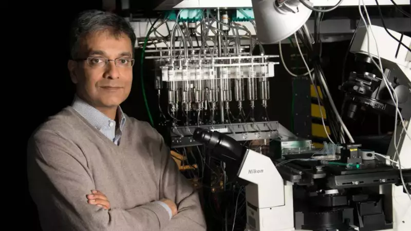
Now, an Indian-origin Harvard professor is pushing this frontier forward. Aravinthan D. T. Samuel, a professor at Harvard University, serves as a senior researcher behind SmartEM. This machine learning-guided system aims to make detailed brain scanning faster, more efficient, and less costly than ever before.
Who Is Aravinthan Samuel?
Samuel has built his career at the intersection of physics and brain science. He uses quantitative methods to explore how living systems behave. His research focuses on how brains convert information into action. He is widely recognized for work linking biology, computation, and neuroscience. Samuel actively supports efforts that make complex brain research easier to carry out at scale.

SmartEM fits into his wider mission by solving a major practical bottleneck in connectomics. The field struggles with the immense time and cost of imaging brain tissue. Samuel is also known as a triple Harvard alumnus. He earned a BA in Physics, completed a PhD in Biophysics, and conducted postdoctoral research in neuroscience at Harvard.
Over the years, he has received major US research honors. These include the NSF CAREER Award and the Presidential Early Career Award for Scientists and Engineers. He also won the NIH Director's Pioneer Award, which recognizes bold, high-impact scientific work.
What Exactly Is SmartEM?
SmartEM is a method that makes microscopes smarter by using artificial intelligence. The AI decides where the microscope should focus its attention. Normally, scientists use electron microscopes to scan extremely thin slices of brain tissue. These microscopes capture astonishing detail, but they take a very long time to scan large areas. The data they produce is enormous.
SmartEM improves this workflow by scanning in a more strategic way. Instead of treating every part of a sample equally, the system performs a quick initial scan. It identifies areas likely to contain important neural structures. Then it spends more time scanning those specific sections in high detail. The idea is simple: save time and effort without losing the precision required for serious brain research.
Why Does Brain Mapping Matter So Much?
The brain operates entirely on connections. Every movement, memory, emotion, and decision is driven by networks of neurons passing signals. Scientists in the field of connectomics try to map these networks in high resolution. Their goal is to understand how the brain is wired and how information flows through it. They also study what changes when disease or injury disrupts these pathways.
Faster and cheaper brain mapping could accelerate research into several critical areas:
- How neural circuits shape behavior
- How brain disorders alter connectivity
- How different species' brains are organized
- How learning and memory are physically encoded in the brain
Making Advanced Research More Accessible
One of SmartEM's biggest advantages is its potential to reduce reliance on rare, extremely expensive imaging machines. The long-term goal is to help more scientists perform high-level connectomics research. The system aims to make commonly available microscopes capable of work that previously required specialized setups.
If this promise holds true, it could broaden participation in brain mapping. More laboratories could join the effort, which would speed up discoveries across all of neuroscience.
Why This Work Stands Out
The biggest breakthroughs in neuroscience will not come from new theories alone. They will come from new ways of seeing the brain in unprecedented detail. Crucially, they must do this fast enough to make discoveries practical. SmartEM is part of that important shift.

By using machine learning to guide electron microscopy, it represents a move toward research tools that are not just powerful, but also efficient and scalable. For Samuel and his collaborators, the mission is clear. They want to take brain mapping out of the realm of rare, slow, high-cost projects. They aim to move it closer to an everyday scientific tool. More laboratories could then use it to unlock what the brain is really doing beneath the surface.