
The Rise of Silent Heart Disease Among Adults Over 40
Heart disease is no longer a distant concern reserved for old age. Increasingly, it is affecting Indians in their 30s and 40s, posing a significant public health challenge. Cardiologists across the country are observing a steady rise in coronary artery disease among adults over 40, not just in metropolitan cities but also in smaller towns. What is particularly alarming is that many cases remain silent, showing minimal or no warning signs until a major cardiac event, such as a heart attack, occurs unexpectedly.
India's Cardiovascular Burden and Modern Lifestyle Factors
India continues to carry one of the highest burdens of cardiovascular disease globally. National health data consistently identifies heart disease as a leading cause of mortality, with clogged arteries now being diagnosed in individuals much younger than previously expected. This shift is largely attributed to modern lifestyles that accelerate heart disease through various risk factors:
- Long working hours and chronic stress
- Sedentary habits and lack of physical activity
- High-calorie diets and poor nutrition
- Smoking and tobacco use
- Uncontrolled diabetes and high blood pressure
Coronary artery disease develops gradually, with plaque building up silently inside the arteries over time, narrowing blood flow. Symptoms may only appear once the blockage becomes significant. Importantly, chest pain is not always the first sign. Some individuals experience mild discomfort, breathlessness during exertion, or fatigue—symptoms often mistaken for acidity or stress, leading to delayed diagnosis.

The Critical Role of Early Detection and Advanced Imaging
Early diagnosis is revolutionizing how heart disease is managed. Traditional tests such as ECGs and treadmill stress tests are helpful but may not always detect early plaque buildup or moderate narrowing. As a result, cardiologists increasingly rely on advanced imaging techniques that allow direct visualisation of coronary arteries. One of the most significant advancements in this field is 640-slice CT cardiac imaging, which offers enhanced precision and speed.
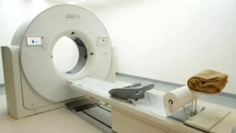
What is a 640-Slice CT Scan?
Unlike conventional CT systems, a 640-slice CT scanner can capture the entire heart in a single rotation. Modern systems operate at imaging speeds of 0.25 seconds per rotation—a crucial advantage since the heart is constantly in motion. The entire heart can typically be scanned within seconds, with the overall procedure completed in under 30 minutes, making it more efficient and less invasive.
Dr. A Sharath Reddy, Senior Consultant Interventional Cardiologist at Medicover Hospitals, explains: This advanced system offers ultra-fast imaging speed, enabling detailed visualisation of coronary arteries within seconds. Because of its rapid capture capability, the need for extensive breath-holding or aggressive heart rate control is significantly reduced. This makes the procedure more comfortable while maintaining high diagnostic accuracy.
Faster imaging reduces motion-related artefacts and enhances clarity, which is particularly beneficial for elderly patients or individuals who may find prolonged breath-holding difficult.
How 640-Slice CT Imaging Detects Silent Heart Disease
A 640-slice CT scan produces high-resolution 3D images of coronary arteries, allowing doctors to:
- Assess plaque characteristics and composition
- Measure the degree of narrowing in blood vessels
- Evaluate calcium deposits and their impact
- Detect early-stage coronary artery disease before symptoms manifest
CT coronary angiography is non-invasive for routine diagnostic evaluation and does not require catheter insertion. Instead, an intravenous contrast injection is used to visualise blood vessels clearly. For adults over 40—especially those with risk factors such as diabetes, hypertension, smoking history, obesity, or a family history of heart disease—early imaging assessment can play a crucial role in preventing major cardiac events by enabling timely intervention.

Availability of Advanced Cardiac Imaging in Hyderabad
Access to advanced diagnostic technology significantly improves early detection outcomes. The 640-slice CT cardiac imaging system is available at Medicover Hospitals in the Financial District, Hyderabad. The availability of such advanced infrastructure enables patients to undergo high-resolution heart imaging locally without travelling outside the region, facilitating easier access to critical healthcare services.
Key Takeaways and Recommendations
Silent heart disease progresses quietly, often with symptoms appearing only when the blockage becomes severe. The important question remains: Is your heart truly safe? With faster imaging, enhanced precision, and improved patient comfort, 640-slice CT technology is emerging as a valuable tool in early cardiac evaluation. Because early detection does not just identify disease—it helps prevent emergencies by allowing for proactive management.
If you are above 40 or have risk factors such as diabetes, high blood pressure, smoking history, or a family history of heart disease, consult a qualified cardiologist to understand whether advanced cardiac imaging is appropriate for you. Taking proactive steps can safeguard your heart health and reduce the risk of life-threatening events.
Disclaimer: The views and opinions expressed in this article are based on independent professional judgment of the experts, and we do not take any responsibility for the accuracy of their views. This should not be considered as a substitute for medical advice. Please consult your treating physician for more details. Sahrudaya Health Care Private Limited is solely liable for the correctness, reliability of the content and/or compliance of applicable laws. The above is non-editorial content and TIL does not guarantee, vouch or endorse any of it. Please take all steps necessary to ascertain that any information and content provided is correct, updated, and verified.