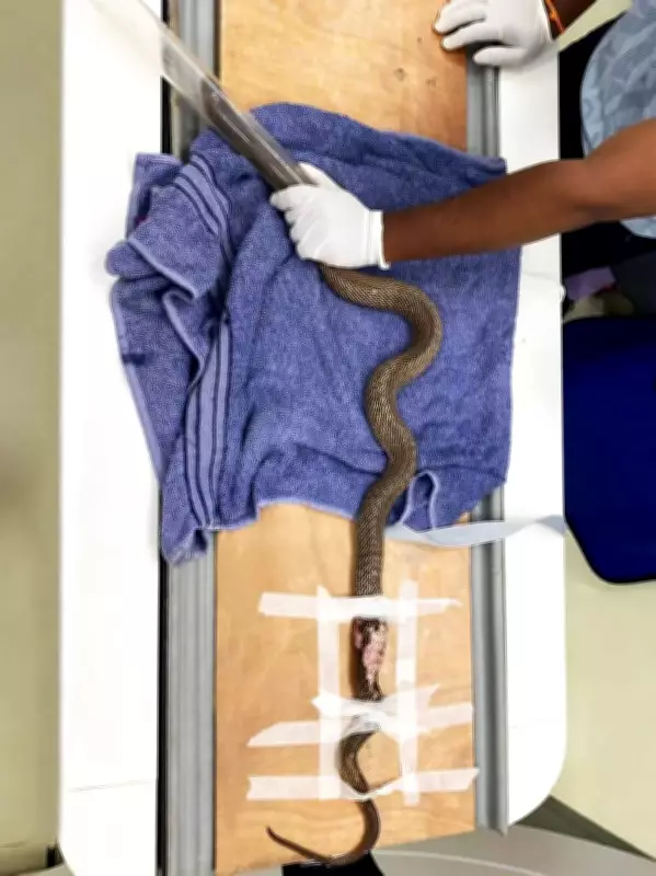
Navi Mumbai: An adult Indian Spectacled Cobra, which was injured in its midsection at a Chembur construction site, underwent a rare and successful CT scan at Fredna Vet Diagnostics (FVD) to fully treat its injuries, a team member said.
Rescue and Treatment
The snake was referred following a history of trauma by veterinarian Dr Deepa Katyal. After recovering, the cobra was released in the wild.
Challenges During the Scan
It was a challenge to ensure that the cobra remained motionless during the CT scan. A team member of FVD said the scan marked a significant step in the use of cutting-edge diagnostic imaging in exotic and wildlife veterinary care.

Procedure Details
A detailed plain CT examination of the caudal coelomic region was conducted at FVD using high-resolution thin-section imaging with multiplanar reconstruction techniques.
Given the inherent challenges of imaging a live reptile, a targeted local nerve block was administered to the cobra before the procedure. This allowed the serpent to be safely and humanely immobilised and taped in position, without the need for general anaesthesia.
Scan Findings
The scan revealed:
- A fracture involving a right-sided rib in the mid-to-caudal coelomic region with mild displacement
- Multifocal subcutaneous air pockets along the body wall
- Soft tissue irregularity and thickening correspond to the external traumatic lesion
Importantly, the CT scan showed no evidence of major organ compromise or vertebral damage, enabling veterinarians to better assess the extent of trauma and plan appropriate treatment and rehabilitation.
Expert Comments
“This case demonstrates the growing role of advanced imaging in reptile and exotic animal medicine. CT technology allows us to evaluate delicate anatomical structures with remarkable precision, especially in challenging wildlife trauma cases,” said a member of Fredna Vet Diagnostics Team.
Stay updated with the latest Mumbai news. Download the TOI App.